An innovative tool used in this research is Digital Image Processing
(DIP) (for our previous applications in human paleobiology and paleoanthropology,
see Macchiarelli et
al., 1990, 1995,
1996a,
b,
1997a,
b;
Bondioli et al.,
1993; Macchiarelli
and Bondioli, 1994, 1995;
Geusa et al.,
1995, 1996a,
b, 1997a,
b; Galichon
et al., 1996; Rossi
et al., 1996, 1997a,
b,
c;
Salomone et al.,
1997; Savorè
et al., 1997; see also Liebermann
et al., 1990; Pesce
Delfino and Lettini, 1990; Potente
et al., 1991, 1992).
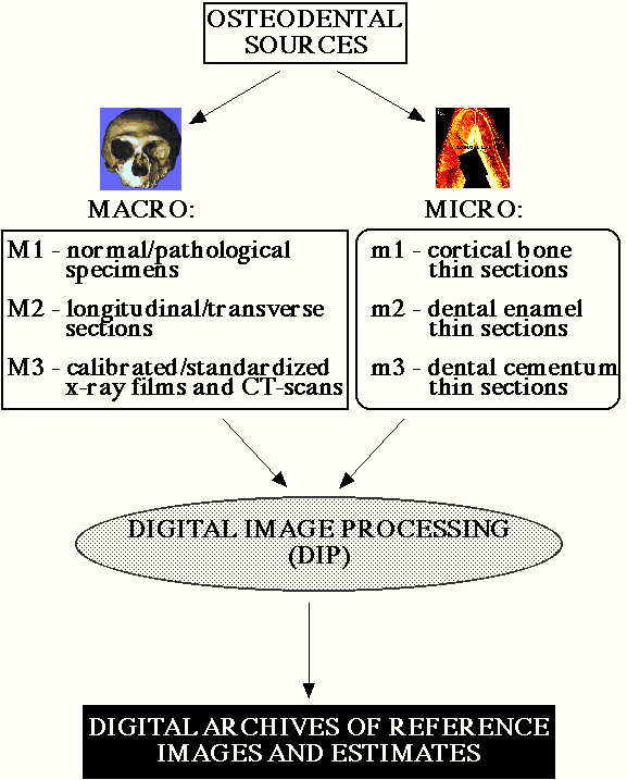
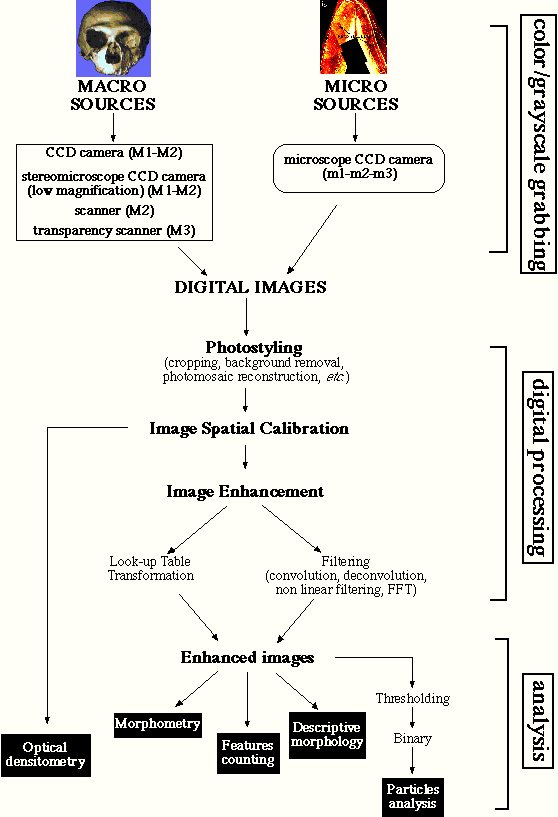
This approach permits a significant enhancement of the quality and quantity
of (histo)morphological, (histo)morphometric, and densitometric information
directly or indirectly obtainable from osteodental remains. By means of
DIP procedures, it is possible to produce a detailed record of (micro)structures
that would otherwise be difficult to obtain and, above all, would produce
highly subjective results (Rosenfeld
and Avinash, 1982; Watkins
et al., 1993).
Each section was first observed at 100x in order to evaluate the presence
and the extension of Wilson bands and their position on both buccal and
lingual aspects of the tooth. Prism bending, as well as the neonatal line,
were observed at a higher magnification (400x).
In order to enhance contrasts, each magnified thin section picture was
transferred into numerical format by means of a digital image analysis
system.
The slices were examined with an optical
microscope (optical transmitted light microscope Laborlux
S, Leica AG) under polarised light and using the Leica l
filter. The microscope is equipped with a
high resolution CCD video camera (TK 1281 Colour Video Camera, JVC Ltd.)
connected to an analogue-to-digital (A/D) converter board (Image Grabber
IG/PC, Neotech Ltd.) plugged into a Pentium PC. The PC is linked with
a mixed network of computer workstations
(PC Pentium, Apple Power Macintosh, Indigo Silicon Graphics). This computer
network has been designed in order to maximise performance in terms of
digital image processing power and versatility. The network design mixes
different software/hardware workstations that are able to run the majority
of modern digital image analysis software. This tool set, combined with
a large on-line storage facility (greater than 20 Gb), guarantees productivity
and efficient use of time during all phases of data collection.
The portion of the thin section covered by the microscope field at 40x
magnification (4.8 mm2) was shot by means of the camera connected
to the microscope, and the resulting analogue image was converted into
numerical format through the image grabber board.
The software (Image Grabber PC 1.01, Neotech Ltd.) that drives the board
returns the final digital image as an average of 16 - or more, according
to user choice - subsequent shots of the same field. This iterative procedure
was employed in order to filter and reduce the noise coming from the CCD
device and from signal deterioration. The spatial resolution was 6.62 mm
for each image pixel, and each final image was stored as an 8 bits, greyscale,
GIF file.
To estimate neonatal line thickness (after Eli
et al., 1989), three images were taken at 400x in
three different positions along the buccal aspect: 1) close to the dentino-enamel
junction; 2) in the middle of the dental crown; and 3) close to the apex.
At 400x magnification the spatial calibration coefficient was 0.66 mm
for each image pixel.
The spatial calibration coefficients were derived from a series of images
- shot at various combinations of magnification and image size - of a micrometry
slide (Leica AG) bearing an engraved metric scale
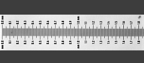 (40x magnification
factor)
(40x magnification
factor)
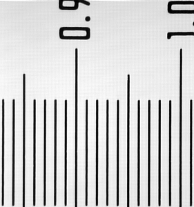 (400x magnification
factor)
(400x magnification
factor)
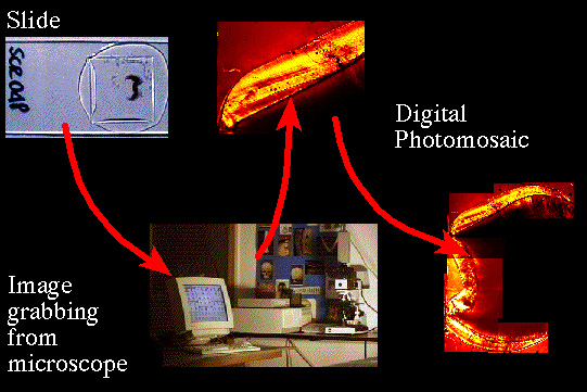
Each image covered only a single portion of the tooth crown section,
thus the final image of the whole crown is the result of the digital
photomosaic of 7 to 15 partial images,
depending on individual tooth size. In order to standardise the images
and to keep the instrumental parameters constant, the program setting of
tone and brightness corrections and microscope light were left unmodified
throughout the same thin section. The photomosaics were created using the
computer program Adobe Photoshop 3.2 (Adobe System Inc.).
In order to estimate the age of formation of the microscopic defects,
different techniques of digital image analysis were used.
The software used for processing is a compound of three different DIP
packages: NIH Image v.
1.61 (National Institute of Health, USA); Optilab
Pro 2.5 (Graftek srl); and UTHSCA
Image Tool 1.28 (Univ. of Texas, San Antonio Health Science
Center, USA).
Contrast enhancement convolution filters (3x3 and 5x5 kernels) were
run to get sharper details, while a change in the look-up table function
was performed to increase site-specific contrasts of intensity profiles.
Further image enhancements of the enamel banding was obtained by the
application of directional embossing filters (usually NE and NW 3x3 and
5x5 kernels) capable of enhancing the transition between the dark and clear
regions of a band.
Special care was taken to avoid the introduction of artefacts, especially
during the convolution and embossing procedures. After each electronic
manipulation, the digital results were carefully compared to the original
images, and each new highlighted feature was considered reliable only when
confirmed by using different analytical tools. All measurements were taken
on images that were elaborated as little as possible.
Measurements on the photomosaics were performed using the National
Institute of Health NIH Image v.1.61 program running on
an Apple Power Macintosh. The path of the measurements was traced by hand
with a mouse on a PC high resolution 21" screen using the straight
polyline measurement tool provided by the program. In order to closely
follow curved paths, the number of the polyline vertex was proportionally
increased where the tightness of the curve increased.
As shown by repeated tests for intra- and interobserver concordance,
the systematic use of DIP techniques as described above ensure a high degree
of repeatability and replicability of the estimates derived from the histomorphometric
record.
Cited References
Bondioli L., Sperduti A., Macchiarelli R. (1993)
Analisi quantitativa delle dinamiche architetturali del tessuto osseo umano
in funzione dell'età tramite elaborazione digitale d'immagine. Antropologia
Contemporanea, 16: 27-32.
Eli I., Sarnat H., Talmi E. (1989) Effect of the
birth process on the neonatal line in primary tooth enamel. Pediatric
Dentistry, 11: 220-223.
Galichon V., Bondioli L., Macchiarelli R. (1996)
Trabecular architecture of the hominid pelvis. American Journal of Physical
Anthropology, suppl. 22: 108-109 (abstract).
Geusa G., Arcudi G., Bondioli L., Capucci E., Mauriello
S., Piccirilli A., Macchiarelli R. (1997a) Correlazione tra età
cronologica e anulazioni del cemento dentario umano in un campione di età
nota. XII Congresso degli Antropologi Italiani. Storia del Popolamento
del Mediterraneo: Aspetti Antropologici, Archeologici e Demografici,
Palermo (abstract). Antropologia Contemporanea (in press).
Geusa G., Bondioli L., Capucci E., Condo' S.G., Macchiarelli
R. (1996a) Le anulazioni del cemento dentario umano. Odontostomatologia,
22/5: 672-676.
Geusa G., Bondioli L., Capucci E., Macchiarelli R.,
Salvadei L. (1995) Anulazioni del cemento e determinazione dell'età
alla morte. In (C. Peretto & S. Milliken, eds.) XI Congresso degli
Antropologi Italiani. L'Adattamento Umano all'Ambiente. Passato
e Presente. Isernia: C. Iannone Ed., pp. 140-141 (abstract).
Geusa G., Bondioli L., Capucci E., Rossi P.F., Macchiarelli
R. (1996b) Anulazioni del cemento e determinazione dell'età alla
morte. In (C. Peretto & S. Milliken, eds.) L'Adattamento Umano all'Ambiente.
Passato e Presente. Atti dell'XI Congresso degli Antropologi Italiani.
Forlì: ABACO, pp. 325-336.
Geusa G., Bondioli L., Macchiarelli R. (1997b) Applicazioni
e archivi digitali in paleobiologia umana tra ricerca, documentazione,
divulgazione scientifica. XII Congresso degli Antropologi Italiani.
Storia del Popolamento del Mediterraneo: Aspetti Antropologici, Archeologici
e Demografici, Palermo (abstract). Antropologia Contemporanea
(in press).
Lieberman D.E., Deacon T.W., Meadow R.H. (1990) Computer
image enhancement and analysis of cementum increments as applied to teeth
of Gazella gazella. Journal of Archaeological Science, 17:
519-533.
Macchiarelli R., Bondioli L. (1994) Linear densitometry
and digital image processing of proximal femur radiographs: implications
for archaeological and forensic anthropology. American Journal of Physical
Anthropology, 93: 109-122.
Macchiarelli R., Bondioli L. (1995) Advanced technologies
in human paleobiology: toward a public access to the odontoskeletal collections.
In (C.N.R., ed.) 1st International Congress on Science and Technology
for the Safeguard of Cultural Heritage in the Mediterranean Basin.
Catania: Litostampa Idonea, p. 383 (abstract).
Macchiarelli R., Bondioli L., Coppens Y., Galichon
V. (1995) L'architettura trabecolare dell'osso dell'anca negli Ominidi.
In (C. Peretto & S. Milliken, eds.) XI Congresso degli Antropologi
Italiani. L'Adattamento Umano all'Ambiente. Passato e Presente.
Isernia: C. Iannone Ed., pp. 45-46 (abstract).
Macchiarelli R., Galichon V., Bondioli L., Tobias
P.V. (1996a) Hip bone trabecular architecture and locomotor behaviour in
South African Australopithecines. In XIII International Congress of
Prehistoric and Protohistoric Sciences. The Sections, Abstracts, vol. 1.
Forlì: ABACO, pp. 109-110 (abstract).
Macchiarelli R., Galichon V., Bondioli L., Tobias
P.V. (1997a) Hip bone trabecular architecture shows uniquely distinctive
locomotor behaviour in South African australopithecines. Journal of
Human Evolution (in press).
Macchiarelli R., Geusa G., Rossi P.F., Salomone
F., Sperduti A., Bondioli L. (1996b) Tecnologie avanzate in paleobiologia
umana: verso un accesso pubblico alle collezioni odontoscheletriche.
In (C.N.R., ed.) Science and Technology for the Safeguard of Cultural
Heritage in the Mediterranean Basin. Catania: Litostampa Idonea (in
press).
Macchiarelli R., Rook L., Bondioli L. (1997b) Reconstruction
of the locomotor behaviour in fossil primates - with special reference
to Oreopithecus - by means of digital image processing of the hip
bone trabecular architecture. XII Congresso degli Antropologi Italiani.
Storia del Popolamento del Mediterraneo: Aspetti Antropologici, Archeologici
e Demografici, Palermo (abstract). Antropologia Contemporanea
(in press).
Macchiarelli R., Sperduti A., Bondioli L. (1990)
L'indagine radiografica dello scheletro nella attribuzione dell'età
alla morte. II. Analisi sperimentale dei corpi vertebrali. Rivista di
Antropologia, 68: 103-127.
Pesce Delfino V., Lettini T. (1990) Elaborazione
di dati da immagini. Giornale Italiano di Ostetricia e Ginecologia,
12: 174-180.
Potente F., Vacca E., Pesce Delfino V. (1991) Valutazione
dello stato di usura dentaria con teniche di analisi di immagine. Antropologia
Contemporanea, 14: 149-156.
Potente F., Vacca E., Pesce Delfino V. (1992) Dental
wear evaluation by image analysis methods. Anthropologie (Brno),
30: 9-12.
Rosenfeld A., Avinash C.K. (1982) Digital Picture
Processing. New York: Academic Press.
Rossi P.F., Bondioli L., Condò S.G., Geusa
G., Macchiarelli R. (1997a) Linea neonatale dello smalto e dinamiche della
nascita: evidenze archeo-istologiche dalla Roma imperiale. Odontostomatologia,
23/5: 546-553.
Rossi P.F., Bondioli L., Geusa G., Macchiarelli
R. (1996) Stress e adattamento in età romana imperiale. In (C. Peretto
& S. Milliken, eds.) L'Adattamento Umano all'Ambiente. Passato e
Presente. Atti dell'XI Congresso degli Antropologi Italiani.
Forlì: ABACO, pp. 343-354.
Rossi P.F., Bondioli L., Geusa G., Macchiarelli
R. (1997b) I microdifetti di sviluppo dello smalto nella dentizione primaria.
Analisi del segmento infantile della comunità romana imperiale del
Portus Romae (necropoli di Isola Sacra) mediante nuove tecnologie
digitali d'indagine. Quaderni del Civico Museo del Finale, 3: 29-38.
Rossi P.F., Bondioli L., Macchiarelli R. (1997c)
Istomorfometria dello smalto dentario in relazione all'evento della nascita.
XII Congresso degli Antropologi Italiani. Storia del Popolamento del
Mediterraneo: Aspetti Antropologici, Archeologici e Demografici, Palermo
(abstract). Antropologia Contemporanea (in press).
Salomone F., Bondioli L., Dazzi M., Geusa G., Pedicelli
G., Sperduti A., Macchiarelli R. (1997) Variazioni strutturali attraverso
l'età dell'osso corticale e trabecolare nelle popolazioni umane
del passato: rilievi morfometrici, radiografici, tomografici ed elaborazione
digitale di immagine. XII Congresso degli Antropologi Italiani. Storia
del Popolamento del Mediterraneo: Aspetti Antropologici, Archeologici e
Demografici, Palermo (abstract). Antropologia Contemporanea
(in press).
Savorè C., Bondioli L., Formenti D., Geusa
G., Grupe G., Rossi P.F., Macchiarelli R. (1997) Analisi archeo-istologica
e processi diagenetici dei tessuti osseo e dentari. XII Congresso degli
Antropologi Italiani. Storia del Popolamento del Mediterraneo: Aspetti
Antropologici, Archeologici e Demografici, Palermo (abstract). Antropologia
Contemporanea (in press).
Watkins C., Sadun A., Marenka S. (1993) Modern
Image Processing: Warping, Morphing, and Classical Techniques. Boston:
Academic Press.